
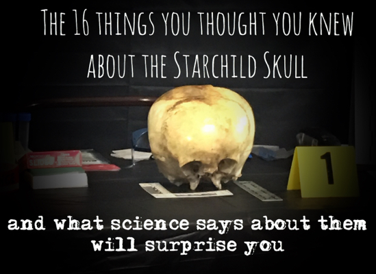
(Warning: Some images in the Investigation Report can be disturbing!)
While Kerry McClure and I, Chase Kloetzke, organized an investigation plan as independent investigators for the infamous Starchild Skull, we ventured forward with the realities and challenges of providing scientific explanations as opposed to the proclamations that have been made previously. Many of these claims either do not have supportive evidence or documented opinion behind them or scientific testing is not possible to explain, confirm or rebuke the anomalies.

So, we made a list. We focused on the claims and visible features that we could tackle professionally, scientifically and, with proper investigation, could produce results with testing that would meet and pass the scientific and public scrutiny. If we could not offer the public every detail on each scientific examination such as the name of the examiner, the place this was done, the work sheets or reports about their process and the actual final report, we would have to reconsider where we focused our time and money. In other words, if we couldn’t test it, prove it or find a valid conclusion, we crossed it off our “To Do” list.
The Field Reports’ findings on reported claims: True or False
1. The Starchild was an adult.
The first decision we made was obvious; we could not compare the two skulls with each other. They are different. Apples and oranges. One is an adult and female and one is most likely on first look and of the opinion of several experts, a child. Many of the many claims previously made make the mistake of assuming the Starchild Skull is that of a fulling grown adult. Of course many of the measurements would seem anomalous if that were true. The age of the Star Child is not in dispute with the team of The Field Reports. We have found only one contradiction of the age previously made from an unnnamed source. Since then, there has been an across-the-board agreement between all experts, including a dentist basing his opinion on his tooth analysis, and lab results that the Star Child is indeed…a child – a 5-year-old child.
Answer: False
2. The eye sockets are unexplainably shallow.
Anyone can clearly see any Starchild Skull photo easily attainable on Google images that the eye sockets are broken. This could greatly affect their appearance and measurements. Conclusive irregularity is also dependent on the age of the Skull. The accepted age from every professional we had examine the skull, found it to be 5 years old, which offers the conclusion of NO irregularity in depth, width, or structure, other than the obvious breaks on the skull. If the argument of shallow eye sockets persists, there is another explanation possible – Crouzon Syndrome. a genetic disorder that prematurely fuses sutures together, causing bulging, wide-set eyes with…shallow sockets. Another possibility is mid-face hypoplasia, which is similar to Crouzon Syndrome, and can be caused by Achondroplasia (see #16). We will wait to see if the DNA tests rules this out.

Answer: False
3. The Skull bone density is thinner than should be.
This was an interesting claim and the images that are found all over the internet does display a significant difference. However, the skulls photographed and compared are of an adult skull and a child – apples and oranges. There is supposed to be this difference. An average adult skull measures 1.25, while a normal young child about the age of 5 should measure approximately 0.5-0.7. Many factors can account for any small variances, especially on the young skull measurements, such as diet and activity levels. The Star Child Skull measured perfectly at 0.7. In other words, when not compared to an adult, the bone density of the Starchild Skull is not anomalous. The Osteology Museum offered many comparison samples that were conclusive to the density AND the weight of the skull.

Answer: False
4. The Star Child Skull displays smaller chewing muscle regions than an average skull.
We can include the following claims because they are all dependent on an adult Starchild opinion, (which has not been the conclusion of any higher-than-normal credentialed scientists and accredited professionals):
- The zygomatic arch is smaller than a normal human.
- The lower and missing parts of the Starchild face will be much smaller than an average adult skull.
The above measurements and anomalies would be under consideration if the Starchild Skull had been concluded as any other age above that of a 5-year-old. Therefore, these claims are not anomalous for a child. Again, every scientific expert that has examined the skull has determined a 5-year-old age. This was also the conclusion of the DNA lab Bold Labs. The team of investigators here at The Field Reports have accepted the professionally unchallenged 5-year-old Starchild conclusion.
Answer: False
5. The ears on the Starchild Skull are considerably lower than should be and the hearing region is twice as big as a normal skull.

The ear placement has been called into question (well, many questions!) and as we saw for ourselves at the Osteology Museum where we had many comparison samples of the appropriate age, a Down Syndrome 5-year-old skull displayed the low ear placement as well. The Field Reports Team has determined that this alleged anomaly is a claim that cannot be proven one way or another. There are no tests completed at this time that could shed proof or even light on the claim. We sidelined this examination…for now. Perhaps, the DNA testing could show whether or not Down Syndrome was a cause for the low ear placement.
Answer: True/Inconclusive
6. The Starchild Skull has no pores – or lucunae.
A normal skull has what is known as lacunae. Our bones are a living tissue that is constantly restoring themselves. These pore-type holes are for the new cell delivery. The Starchild Skull has none of these visible pores:

There is a clear and present difference here, but there is a factor that affects the image; the Starchild Skull has been cleaned, polished and varnished. Yes, lacquered. It would be impossible to rule out the actions that would indeed change the surface of the outer layer of the skull. The SEMs tests that were conducted also show these pores are present inside the layers of bone, so we must assume the pores were present at one time.
Answer: False
7. The dip in the superior or top part of the skull is a result of an abnormally developed sagittal suture.

There is a defined dip in the top part of the Starchild Skull, and could possibly be explained by what is often found with brachycephaly – when this occurs because of the widened bone structure or “short skull symptom”. Before we can determine any cause for the cephalic shape of the Starchild Skull, we must rule out genetic and rare conditions associated with this form. The Field Reports Team does not rule out hydrocephalus as a cause but does go on the record that this condition is a small percentage of the cause. We do not jump to this explanation but it will remain a very low consideration.
Information on comparable cephalic disorders that can cause the “dip”:
The following photo was taken in a museum in Peru:


- This picture illustrates the 2-sided “bulb” formation in Brachycephaly that will produce a “dip” in the middle of a skull affecting the sagittal suture.
Brachycephalic Definition: Having a head nearly as broad from side to side as from front to back, especially one with a cephalic index (CI) over 80.
- CI > 90%, short skull, occiput flattened, widened
- May affect parietal, temporal, and/or frontal bones and facial symmetry
- May occur alone or in combination with plagiocephaly
- May result from the premature fusion of the coronal or lambdoid sutures
Answer: Inconclusive
8. The expanded sides of the skull and extreme flat back of the skull are mysterious.

This is again, a known growth pattern for the Brachycephalic form of a skull, especially when paired with the “Flathead Syndrome” as seen below. This flattened head is a common condition caused by too much time on the back as infants. If the Starchild was disabled, laying on his back, immobile for long periods of time could have contributed to the strange shape of the Skull. It is treated today with a helmet that promotes a round and healthy growth of the skull with small children. Cranial deformation is still an active practice but now it used for corrective procedures.


Answer: False
9. The Starchild Skull has never had frontal sinuses.

The Starchild Skull shows no sinuses compared to the adult. Much more study must be done on the difference of juvenile sinus bone structure compared to a that of a full-grown adult. We do know that juvenile bones are softer and not as rigid as those of an adult and this could be cause for the bone/cartridge parts to be missing. The decay rate of human remains will always depend on depth of burial, soil conditions, and the environment. The narrative of how the skulls were found describe the companion skeleton on the ground and that of the Starchild buried under a mound of dirt. Also, the age of the human is a factor in decay rate; children will have a faster decline. If the Starchild Skull and the companion skull were indeed laid to rest at exactly the same time, exactly the same way in the exact location, we would be able to justify an anomaly, but at this time, no evidence exists that could be scientifically tested to determine if sinuses existed at all.
Answer: Inconclusive
10. The maxilla contained 3 rows of teeth.
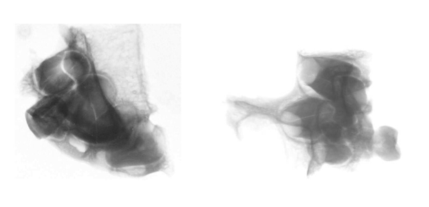
The small piece of the maxilla was found in the bottom of the box when the owner, Melanie Young was given the skulls. It does, in fact, belong to the Starchild. Dr. James Gilliam, DDS. Oklahoma City, Oklahoma, was given the photos and X-rays to examine. His tooth analysis concluded that teeth and tooth buds were what you expect to find on a young child, approximately 5 years old. The “tooth buds” are forming as he reaches the age to lose the baby teeth. Adult teeth do not form completely until they are ready to be exposed and this is what will cause the loosening of the baby teeth. There has not been any professional on record to claim there are 3 rows of teeth. This “tooth” has caused the only controversy in the actual age of the Starchild as one unnamed “expert” (?) concluded the wear on the tooth was more of an adult status. Unfortunately, we cannot verify this proclamation. But all experts and testing since have concluded the deposition as an approximate age of 5.
Answer: False
11. The exterior occipital protuberance (inion) is missing.
There have been two separate expert opinions about this claim as it is a difficult call. The inion can be located but it is so very small, it could also be another anomalous part of the skull. The inion is responsible for a neck muscle attachment (trapezoid). If this small protrusion is indeed the occipital protuberance, this would lend more credence to the theory that the Starchild was not as mobile as a healthy child. The muscles would have not developed as normal, therefore leaving little need for a prominent inion. Bones will strengthen with activity and with weight-bearing functions, therefore, his under-developed part of anatomy could lend corroboration to “Flat Head Syndrome” as seen in children today.


Answer: Inconclusive
12. The head boarding theory
There are no visible or normal indications found on the Starchild Skull that have been documented or examined on thousands of skulls known to be shaped by cranial deformation. The banding marks are noticeably missing. Although this practice cannot be 100% ruled out, there are NO reasons or evidence to found to support head boarding except the shape of the skull which can be explained by other factors.
Answer: False
13. The “fibers” and “red residue” gave the skull above average strength.
SELEE Lab conducted the SEMs testing with the sole purpose of an elemental analysis and to find the reported “fibers”. No fibers were found. None. It is important to remember that the Starchild Skull has been handled by hundreds of people, stored in boxes, wrapped in material for transport, lacquered, and a host of other events. The collective opinion of the fibers is: they are contamination particles that were not removed before varnishing the exterior of the skull. As the varnish soaked in, it embedded the dust and debris into the pores of the bone causing this isolated effect.
It was after the Field Reports lab testing that we learned other earlier tests were done to corroborate the first findings and all testing since has not been able to locate the fibers. ANY fibers. The red residue has also been declared as a contamination spot as it too has not been located on the skull since the original examination and claim. It is hard to determine if there are more fibers and residue because of the damage afflicted on the skull every time a test is run. What we can determine with certainty, is that neither the fibers nor the residue are a continuous anomaly causing any extra strength or abnormally.
Answer: False
14. The DNA proves the Starchild Skull is an alien hybrid.
First of all, if DNA testing on the Starchild showed something so unknown that it may, in fact, be part extraterrestrial, there needs to be a comparison sample to prove that it is part alien. Unfortunately, that does not exist. There has been DNA testing so far:
Bolds Lab
In 1999, Bold Lab, (Bureau of Legal Dentistry) in Canada was contacted to do the DNA test on the Starchild Skull. Two samples were tested. They reported “conclusive evidence” that the Starchild was male and human. As both chromosomes are present in males, X and Y, both parents must be human. The Y chromosome can ONLY be inherited from the Male or Father.
Trace Genetics
In 2003, Trace Genetics, a lab that specializes in extracting ancient DNA concluded the Starchild Skull and the Companion Skull were not related as each belong to a different Haplogroup. The Starchild’s Mitochondrial DNA (inherited exclusively from the mother) has origins in Haplogroup C. The Companion Skull has origins in Haplogroup A and is female. The Companion Skull is NOT related to the Starchild Skull. Both Haplogroups are Native American.
Dr. Garry Nolan PhD. Geneticist/Stanford University
“The facts are not open to interpretation unless they want to rewrite the rules of genetics. There is no analysis available by which they can take the data they have provided me, or on the web, to make the claims they do about FoxP2. I can only guess at the reasons they want to keep up such an obviously incorrect claim…DNA tests have shown it is human (and both parents were human) suffering probably from congenital hydrocephalus.”
Un-named Geneticist
Controversial at best. There is no room in any investigation to reply or even to repeat statements or proclamations from people who will not allow their name, work place, and credentials to be published. The Field Reports Team does know the identity but have promised confidentiality to the Starchild Project on this matter. And ONLY on this matter. We will always honor our word. What we can reveal is that this “geneticist” is NOT a geneticist but a virologist that has worked on Lyme Disease. Maybe this explains the silence.
Answer: False
15. The Debunking Theory: Hydrocephalus
This theory cannot be ruled out nor ignored. The pictures are a better comparison view that a hydrophilic anomaly can and usually does appear symmetrical. There are hundreds of images on the internet and some show extreme and devastating deformities caused by the condition. It doesn’t seem a fair comparison to only show rare cases of extreme hydrocephalus to rule out that as a cause of the Starchild’s skull shape. However, the normal deformity is shown below. We did not include the toddlers nor the young children’s images as they are often photo shopped; meaning, their eyes are blacked out, faces blurred and we saw no reason to go any further with these already heartbreaking images. We did have to offer a better sample pool of what hydrocephalus looks like in a “typical” state.


The photos illustrate a better comparison to a Hydrocephallic condition.
DNA cannot test for this condition. We cannot find a test for this. This possibility must stay in an honest assessment when considering the larger cranial capacity that is NOT in dispute at this time. The skull is bigger. Normal head boarding or cranial deformation does not increase the cranial capacity which is another corroboration that the Skull was NOT cradle boarded. Upon further research, the list of similarities between a child with hydrocephalus and the Starchild is growing. Notice the low placement of the ears caused by hydrocephalus (see #5).
Answer: Inconclusive
16. The foramen magnum is abnormal.
The Starchild Skull’s foramen magnum, or opening at the bottom of the skull is very strange-looking and not placed where a typical skull’s opening is. There is a possible explanation for this – Achondroplasia. This is a genetic mutation on the FGFR3 gene that causes various disorders and symptoms including:
- dwarfism
- larger than average head size (check!)
- Cervico-medullary Myelopathy which causes a compression in the foramen magnum (foramen magnum stenosis or having a smaller than average foramen magnum is one of the main symptoms) and this causes the spinal cord to kink, possibly causing motor regression, or immobility which would lead to “flat head syndrome” if the child were left on its back (see #8)
- hydrocephalus (see #15)
- mid-face hypoplasia (see #2) which causes shallow eye sockets
- anatomical problems with the ears due to the compression of the brain stem at the foramen magnum (see #5)
- defective enchondral growth which affects the creation of bones and can cause deformities (the skeleton of the Starchild was reported to be deformed by the young girl who found it).
- basilar invagination – where the tip of the cervical vertebrae exends into the foramen magnum – often associated with platybasia – or the flattening of the base of the skull. Could this be the cause of the strange shape of the Starchild’s foramen magnum?
Answer: True/Inconclusive
The Final Say
The Field Reports is not here to give a final verdict, but to present the facts to date. All we can do is offer all of the research and observations made by professionals so that we and you can form a more educated guess, or at least dig deeper when reading other’s claims. Our thorough research currently points to a possible natural reason for the anomalies with Achondroplasia (which can be proven or ruled out with DNA) or something similar looking like a good candidate, or at least hydrocephalus. Perhaps the DNA testing being conducted right now will give us a more conclusive answer to these questions and more. Of course, it would be so exciting to have results so unexplainable that further study would be necessary, but until then, we will wait. We encourage you to weigh in on our findings so far. If you happen to be an expert in congenital birth defects we would really appreciate your input and if you would like to join the discussion, please leave a comment or join our Facebook page.
Stay tuned as we are working on more brand new information pertaining to the Starchild’s origins!

1 Pingback